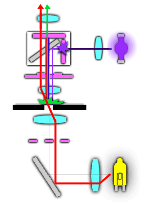
Fluorescent � and Phase Contrast Light Path Combined
�
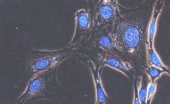
There may � be times when it is useful to locate and view a specimen using phase contrast � and then add fluorescence illumination to view an organelle or the location � of a particular molecule in the cell. Once the specimen, say a cell, is � located under phase contrast, the light intensity knob is used to turn � down the halogen lamp and the fluorescence shutter is set to the "on" � position. The correct filter cube is placed in the light path and the � intensity of the halogen lamp is adjusted so that the cell can be seen � (with phase) and the fluorescing structure(s) is/are visible (see the � figure below).
�
A
�
cell viewed by phase contrast with a blue
�
fluorescent nucleus stained with DAPI
�
(courtesy Olympus Microscope Co.)
