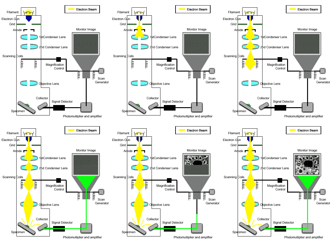
Electron Beam Path

© 2003 Wartburg College Biology Department

BIOLOGY DEPARTMENT HOME PAGE

Scanning Electron Microscopy (SEM)

The scanning electron microscope utilizes a focused beam of high energy electrons that scans across the surface of the sample. As the beam interacts with the sample it produces a large number of electrons at or near the surface. In the SE mode, almost all the low energy (secondary) electrons and some of the "backscatter" electrons are collected at a positively biased collector. In the NSEM mode, the SE collector is shut off which allows for detection of only the higher energy (but weaker detection signal) "backscatter" electrons using a Robinson detector.

In either case, the scattered electrons strike a scintillator on the collector which contains phosphors that produce small flashes of light (photons). These photons are converted to an amplified electronic signal that displays on a cathode ray tube (CRT, similar to a TV tube). The scanning of the beam across the sample is synchronized with the scanning on the CRT, so you have a one-to-one relationship between points on the sample and points on the screen.

The contributions of the SEM to science comes from its ability to combine high magnification (routinely up to 100,000x) with great depth of field. It also bridges the magnification gap between the light microscope and the transmission electron microscope.

BACK TO VIRTUAL SEM HOME PAGE