
Fluorescent � Light Path
�
�
The fluorescent � microscope is used to visualize specimens that fluoresce, that is to say, � emit light of a different color (always a higher wavelength) than the � light absorbed by the specimen. Fluorescence occurs either because of � naturally occurring fluorescent materials in the cells (such as chlorophyll � and related molecules) or because the cells have been stained with a fluorochrome. � Fluorochromes are stains similar to cell and tissues stains used in light � microscopy and have been chosen or designed to be highly specific in their � attachment to molecules in cells. The use of fluochromes has made it possible � to view cells and subcellular components of cells with a high degree of � specificity amid non-fluorescing material (see the DAPI stained nucleus � below).
�
� �
A cell nucleus � stained with DAPI (courtesy Olympus Microscope Co.)
�Each fluorochrome
�
has a specific excitation wavelength and emission wavelength and there
�
are many fluorochromes in use today. Thus, fluorescent microscopes are
�
generally equipped with several types of fluorescent filter cubes. The
�
most common cubes are labeled as B, G and U filter emitters. Link
�
to Applications of Fluorescent Microscopy for more detail on filter cubes.
�
�
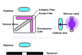
FLUORESCENCE LIGHT PATH
The animation � at the left shows the optical system for an epi-fluorescence upright microscope. � The mercury lamp emits ultraviolet (UV) light that is directed to a specific � fluorescence filter cube located in the mirror turret unit. The exciter � filter in the cube is designed to allow specific wavelengths of UV light � to pass though (those that are absorbed by the dye in use) which are reflected � by the dichroic mirror and pass through the objective lens to illuminates � the specimen (violet rays).
�The light � emitted from the specimen (multiple � color rays) is directed upwards through the objective and pass through � the dichroic mirror (which removes the incident UV light and allows the � longer wavelength visible light to pass through). The light emitted is � viewed after passing through the emission filter which selects which wavelength � of light passes through to the eyepieces (only the green � ray). In this case, one sees a bright green glowing dye-stained cell or � organelle against a dark background.
�