Phase
�
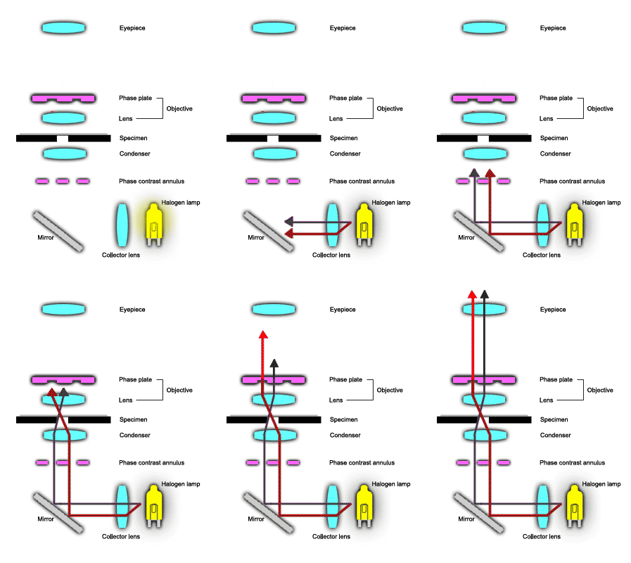
Contrast Light Path
�

WHY PHASE � CONTRAST MICROSCOPY?
�Living cells � and most cell organelles are often difficult if not impossible to see � by brightfield or darkfield microscopy because they do not absorb, refract � or diffract sufficient light to contrast with the surrounding medium. � While staining of specimens with preferential stains (methylene blue for � example stains nuclei blue) have allowed researchers to examine cells � structures, staining generally kills the cells and in some cases distorts � the cell structure.
�The phase
�
contrast microscope was developed to improve contrast differences between
�
cells/organelles and the surrounding medium, making it possible to see
�
cells/organelles without staining. Thus we can view wet mounts of living
�
cells.
�
PHASE � LIGHT PATH
�Light rays
�
passing through a transparent specimen emerge as either direct rays or
�
diffracted rays. In phase microscopy, this effect is amplified by using
�
a microscope equipped with a special annulus (below the stage) and phase
�
plates (located in the objectives) which accentuate the phase changes
�
produced by the specimen. Direct rays, unimpeded by the phase plate (red
�
ray) are of higher intensity, making the background bright and the diffracted
�
rays (black ray)
�
impeded by the phase plate are of lower intensity, making parts of the
�
specimen darker. This results in improved contrast differences between
�
the specimen and the surrounding medium, making it possible to see cells
�
and cell organelles without staining.
�
